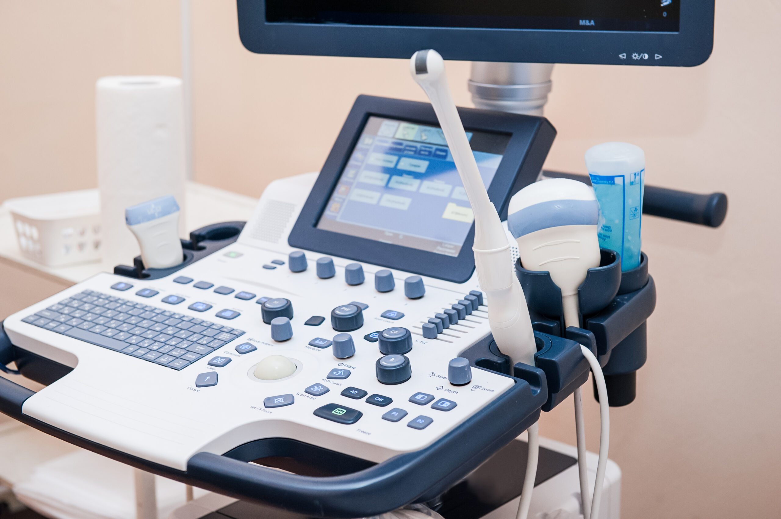
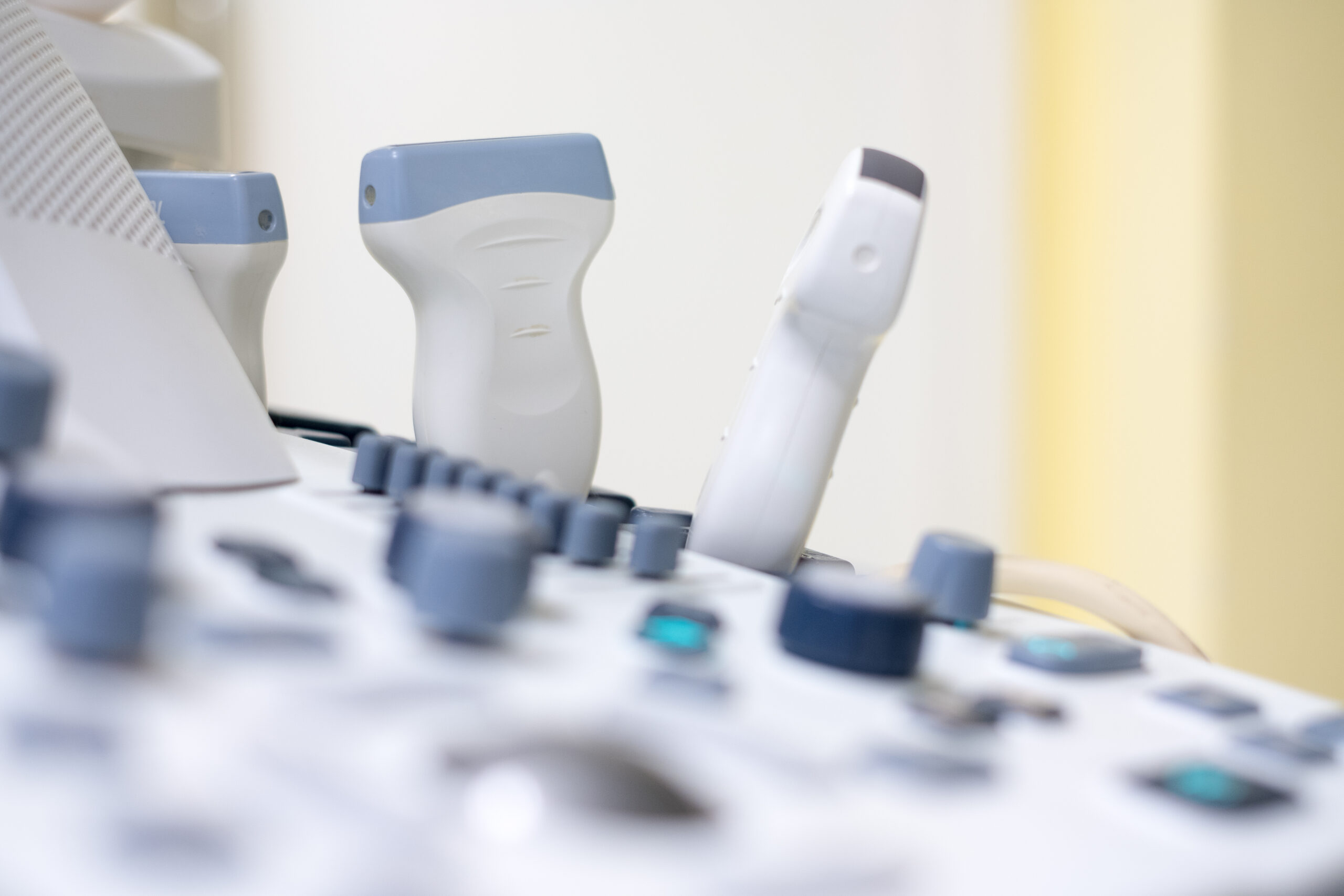
Ultrasonic
Modern ultrasound devices allow you to examine your internal organs painlessly and in detail.
Typical indications for ultrasound examination
- Breast examination
- Examination of the internal genital organs
- Examination of child development during pregnancy
- Exclusion of childhood malformations
- Examination of the thyroid lobes
- Examination of the abdominal organs such as liver, kidneys
This visual representation allows us to quickly make a diagnosis and, if necessary, initiate treatment. Ultrasound is also used for many general medical issues and for the targeted blocking of specific nerves in pain management.


Vaginal sonography of the internal female genital organs makes it possible to detect small changes and abnormalities early on. This ultrasound method allows for detailed visualization of the ovaries and uterus. Accordingly, regular ultrasound examinations can differentiate between benign and malignant diseases of the female genital organs in their early stages.
We recommend routine vaginal ultrasound, especially for women with a family history of the condition or other risk factors. Regular examinations play a crucial role in improving early detection and initiating timely treatment.
Our experienced team will support you in taking the necessary diagnostic measures and developing a therapy tailored to your individual needs.
Breast cancer remains the most common cancer in women, but men are also affected. In addition to breast self-examination, breast ultrasound has gained significant importance in breast cancer screening in recent years. It is particularly suitable for the evaluation of palpable breast changes and is also used in routine screening. Occasionally, further evaluation with mammography is necessary.
Breast elastography is a special ultrasound examination that detects hardened tissue and measures the density of the lesion. It is an optimal and non-invasive complement to the evaluation of initially abnormal findings on palpation. This examination can further support early breast cancer detection.
How does it work?
Pressure from the ultrasound probe deforms the glandular tissue of the breast in different ways. This allows differences in elasticity to be visualized and quantitatively measured.
What is being measured?
The stiffness or elasticity of the tissue may indicate tumors or other pathological changes.
indication
Elastography complements the classic breast ultrasound examination (mammary ultrasound) in cases of unclear findings, e.g. in cases of dense glandular tissue or voluminous breasts.
Why?
The examination makes it possible to obtain further information in practice and without any additional stress, e.g. through X-rays, to distinguish between benign and potentially malignant changes.
Color Doppler sonography is a specialized ultrasound examination method for visualizing blood vessels and measuring blood flow. It is used in gynecology and prenatal care to measure blood flow velocity in vessels. This makes it possible to assess the fetal oxygen and nutrient supply and monitor high-risk pregnancies.